
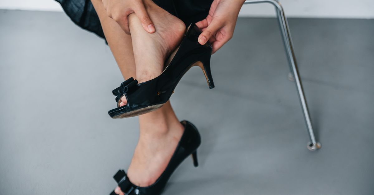
Heel fat pad syndrome occurs when the thick cushion of fatty tissue beneath the heel bone thins, migrates, or loses its shock-absorbing capacity, causing deep, bruising pain directly in the center of the heel. Unlike plantar fasciitis, which typically produces sharp pain at the inner heel that is worst with the first steps in the morning, heel fat pad syndrome produces a diffuse, aching pain that worsens with prolonged weight-bearing and improves with rest. It is most common in older adults due to age-related fat pad thinning, but also affects runners, people who work on hard surfaces, and those who have sustained direct heel trauma. Management focuses on load reduction, cushioned footwear, heel cups, and supportive soft-tissue care rather than aggressive deep-pressure treatment directly on the thinned pad.
The bruised feeling under your heel that gets worse the longer you stand — and doesn't ease up after a few minutes of morning movement — is not the same as plantar fasciitis. Heel fat pad syndrome is a distinct condition, and misidentifying it leads to the wrong treatment plan. People who stretch their arches, ice their fascia, and wait for morning stiffness to pass often find no relief because their actual problem is a degraded shock absorber, not an inflamed ligament.
Heel fat pad syndrome is frequently overlooked, underdiagnosed, and overshadowed by the far more publicized plantar fasciitis. What follows explains exactly what it is, how to tell it apart from other common causes of heel pain, who is most at risk, and what an effective care approach actually looks like — including an honest explanation of where massage helps and where it doesn't.
What Happens to the Heel Fat Pad
The heel bears a significant portion of your body weight with every step. Protecting the calcaneus — the large heel bone — is a specialized structure called the heel fat pad: a complex network of sealed fat chambers reinforced by fibrous tissue. This pad is not simply a layer of general body fat. It is architecturally designed for load absorption, compressing under impact and rebounding between steps.
The Structure of the Pad
The heel fat pad is organized into microcompartments — small columns of fat encased in elastic connective tissue. This structure allows it to compress significantly under load while returning to shape quickly enough to absorb the next footfall. Healthy heel fat pads can be 18 millimeters or more thick and absorb a substantial portion of the impact generated during walking and running.
As this tissue ages or sustains repeated trauma, the fibrous walls between compartments break down. The fat itself may thin, or it may remain present but shift forward and away from directly beneath the heel bone — a condition called fat pad displacement. Either way, the calcaneus ends up bearing more direct impact force than it was designed to handle.
How the Pad Degrades Over Time
Age is the most consistent driver. Research in clinical foot and ankle literature has documented measurable reductions in heel fat pad thickness beginning in middle age, with more pronounced thinning in older adults. The fibrous septa that hold the fat chambers in place gradually lose elasticity, and the pad's ability to rebound between steps diminishes.
Several factors accelerate that degradation:
- Prolonged walking or standing on hard surfaces such as concrete or tile
- High-impact athletic activity, particularly long-distance running with inadequate footwear
- Direct trauma to the heel — a fall, a hard landing, or a single heavy impact
- Corticosteroid heel injections, which can break down local fat tissue over time
- Low body mass index, which correlates with reduced fat pad volume
- Systemic conditions including rheumatoid arthritis and connective tissue disorders
The result is a heel that can no longer adequately cushion each step — leading to pain that accumulates with use rather than clearing with movement.
Why It Gets Confused With Plantar Fasciitis
Both conditions produce heel pain in adults, and both worsen with time on your feet. The similarities end there. Plantar fasciitis is an overuse injury of the plantar fascia ligament, which attaches at the inner aspect of the heel bone. Its hallmark is sharp, stabbing pain at the medial heel that is worst with the first steps after sleep or prolonged sitting — the "first-step pain" phenomenon — and often eases after several minutes of walking as the fascia warms up.
Heel fat pad syndrome produces a different pain profile. The pain is located at the center of the heel, directly underneath the calcaneus rather than at its inner edge, and is described more often as a deep bruise than a sharp stab. It does not follow the morning-stiffness-then-improvement pattern. Instead, it worsens progressively through a day of standing or walking and is relieved by sitting or removing weight from the foot. Walking barefoot on hard floors is often intolerable. These distinctions matter because the treatments differ significantly.
Circulation Support and Soft-Tissue Care
There is no intervention that directly rebuilds a degraded heel fat pad — and that's an important reality to state plainly. The goals of conservative management are to reduce the load on the thinned pad, support the health of surrounding tissue, and keep blood moving through the foot. Circulation supports the connective tissue and musculature that indirectly stabilize the heel.
Where Massage Helps — and Where It Doesn't
Direct, deep pressure applied to an already-thinned heel pad is counterproductive. The pad has already lost its ability to absorb force, so pressing hard into the heel bottom does not stimulate regeneration — it compresses tissue that cannot rebound effectively and can increase soreness. Anyone recommending aggressive deep-heel massage for this condition is treating it like plantar fasciitis, which it is not.
Soft-tissue work on the surrounding structures is a different matter entirely:
- Calf musculature: Tight calves increase the load transferred to the heel during walking. Loosening the gastrocnemius and soleus reduces that downstream force.
- Plantar intrinsic muscles: Gentle work along the arch and ball of the foot supports the broader foot structure without loading the heel directly.
- Achilles and lower leg: The Achilles tendon connects directly to the calcaneus. Improving blood flow in the lower leg tissue around this structure supports overall heel mechanics.
For overall foot circulation, a therapeutic foot massager used at a gentle setting can support blood flow through the lower leg and foot without placing direct downward pressure on the heel. The key is placement and intensity: gentle settings, with the heel positioned to benefit from calf activation rather than direct pad compression.
The Role of Calf Work in Heel Load Reduction
The calf muscles — particularly the gastrocnemius and soleus — play a direct mechanical role in heel loading. When these muscles are tight or fatigued, the heel absorbs more impact force per step because the ankle cannot move through its full range of motion efficiently. Keeping calf tissue supple and well-circulated is one of the more practical things a person with heel fat pad syndrome can do between rest periods.
Regular calf stretching, particularly wall stretches held for 30 seconds at a time, is consistently recommended in clinical guidelines for lower-extremity heel conditions. Gentle massage of the calf belly — not the heel itself — supports the same goal. MedMassager's Foot Massager uses oscillating motion to deliver vibration through the lower leg and surrounding musculature, keeping circulation moving without adding concentrated downward pressure to the heel. Using a foot massager with oscillating technology positioned to engage the calf can help maintain blood flow through the connective tissue structures that stabilize the heel.
Footwear, Heel Cups, and Load Management
Because the underlying issue is a loss of the pad's own cushioning, the most direct and evidence-supported interventions involve replacing that lost cushioning externally and reducing how much daily impact the heel absorbs.
Heel Cups and Cushioned Insoles
Viscoelastic heel cups are specifically designed to address fat pad atrophy. Unlike flat foam insoles, heel cups cradle the heel and mechanically concentrate the remaining fat pad tissue under the calcaneus — partially compensating for displacement. They also add a layer of shock absorption that the degraded pad can no longer provide on its own.
Several clinical reviews have found heel cups to be among the most effective conservative interventions for this condition. They are available over the counter and are worth trying before pursuing more expensive options. Key features to look for:
- Silicone or viscoelastic material rather than rigid plastic
- A deep cup design that holds the heel centrally
- Sufficient thickness — at least 6 to 9mm — to provide meaningful shock absorption
- Compatibility with your footwear, since a heel cup that shifts inside a loose shoe provides little benefit
Footwear Choices That Matter
Walking barefoot or in flat, thin-soled shoes on hard surfaces is one of the most common aggravating behaviors for this condition, leaving the calcaneus with essentially no external protection. Footwear selection should prioritize:
- Thick, cushioned midsoles — EVA or similar materials rather than minimalist soles
- Mild heel elevation, which reduces direct impact on the heel pad without the instability of a high heel
- Good overall fit so the heel cup or insole stays correctly positioned throughout the day
- Avoidance of worn-down soles, since compression zone integrity degrades before the upper shows visible wear
Activity and Load Management
Reducing cumulative heel load is not the same as becoming sedentary. The goal is to eliminate or reduce the highest-impact activities temporarily — running on hard pavement, prolonged standing on concrete — while maintaining lower-impact movement. Swimming, cycling, and walking on grass or soft surfaces keep the body active without transmitting the same forces through the heel.
Periods of planned rest after extended time on your feet allow surrounding tissue to recover. Elevating the feet during rest also supports circulation. This is where gentle foot and calf massage, or the use of a therapeutic massager at low intensity, can complement physical rest by keeping circulation moving through the lower leg without adding new mechanical stress to the heel.
Who Is Most at Risk and When to See a Doctor
Heel fat pad syndrome is not exclusive to older adults, though it is most prevalent in that population. Understanding the at-risk groups helps with both prevention and earlier recognition.
Primary Risk Groups
Certain populations face meaningfully higher risk due to the mechanical and biological factors that accelerate fat pad degradation:
- Adults over 40: Age-related thinning progresses gradually and often becomes symptomatic after increased activity or prolonged time on hard surfaces
- Distance runners and jumping athletes: Cumulative impact accelerates fibrous tissue breakdown, particularly in those who train on asphalt or concrete
- Occupational standing workers: Healthcare workers, retail workers, construction workers, and teachers who spend six or more hours daily on hard floors
- People who have received multiple corticosteroid heel injections: Repeated steroid injections for conditions like plantar fasciitis are associated with fat pad atrophy over time
- People with low body weight: Reduced overall fat tissue can mean lower pad volume at baseline
When Professional Evaluation Is Warranted
Most cases respond to conservative measures, but some warrant clinical assessment. See a podiatrist or orthopedic specialist if:
- Pain is severe enough to limit normal daily walking after several weeks of conservative care
- You are uncertain whether the diagnosis is fat pad syndrome, plantar fasciitis, or a stress fracture — all of which can cause deep heel pain
- You have diabetes, peripheral neuropathy, or circulatory conditions that complicate foot care decisions
- Symptoms worsen rather than plateau or improve with rest and cushioning
Imaging — particularly ultrasound or MRI — can measure fat pad thickness and confirm the diagnosis. A confirmed diagnosis matters because it changes the treatment plan: a stress fracture requires immobilization, plantar fasciitis may benefit from more aggressive stretching, and fat pad syndrome calls for the cushioning and load-reduction approach described here.
Frequently Asked Questions
How do I know if I have heel fat pad syndrome or plantar fasciitis?
The location and timing of pain are the most useful distinguishing factors. Plantar fasciitis typically causes sharp pain at the inner edge of the heel that is worst with the first few steps after sleep or prolonged sitting, then improves as you keep moving. Heel fat pad syndrome produces a deeper, bruise-like pain centered directly beneath the heel bone that worsens progressively through the day with continued weight-bearing and does not follow the morning-stiffness-then-improvement pattern. A podiatrist can confirm the diagnosis, and imaging such as ultrasound can measure fat pad thickness to differentiate the two conditions.
Can heel fat pad syndrome heal on its own?
The underlying structural change — thinning or displacement of the fat pad — does not fully reverse on its own, but symptoms can be managed effectively with conservative care. Many people experience significant pain reduction through heel cups, cushioned footwear, and activity modification. The goal of conservative treatment is to externally replace what the degraded pad can no longer provide and reduce the amount of force the heel must absorb daily. Without addressing these factors, symptoms tend to persist or worsen with continued exposure to hard surfaces and high-impact activity.
Is it safe to massage a heel with fat pad syndrome?
Direct, deep pressure applied to the bottom of the heel is not recommended for this condition. The fat pad has already lost shock-absorbing capacity, and aggressive downward pressure can increase discomfort without providing therapeutic benefit. Gentle massage of the surrounding structures — the calf muscles, the plantar arch, and the lower leg — is more appropriate because it supports circulation and reduces the mechanical load transmitted to the heel without compressing the already-thinned pad.
What kind of shoes should I wear if I have heel fat pad syndrome?
Shoes with thick, cushioned midsoles and a modest heel lift are generally preferred because they absorb impact before it reaches the heel bone. Minimalist shoes, flat-soled footwear, and worn-down soles provide little protection for a thinned pad. Adding a viscoelastic heel cup inside your footwear can significantly improve comfort by mechanically centering and supporting the remaining fat tissue beneath the calcaneus. Walking barefoot on hard floors eliminates all external cushioning and should be avoided.
Does heel fat pad syndrome ever require surgery?
Surgery is rarely necessary and is not a standard treatment for heel fat pad syndrome. The condition is generally managed conservatively through cushioning, load reduction, and supportive footwear. In cases where fat pad displacement is significant, some clinicians have explored injection-based approaches to support pad volume, but these are not widely established. If conservative measures fail after several months and pain significantly limits function, a specialist consultation is appropriate to discuss what options may be suitable for the specific presentation.
Why does walking barefoot hurt so much with this condition?
The heel fat pad normally absorbs the impact of each footfall before it reaches the calcaneus. When the pad has thinned or displaced, that protective layer is diminished, and walking barefoot on hard surfaces transmits far more direct force to the heel bone than it can comfortably tolerate. Even a brief walk across tile or hardwood flooring can produce significant pain because no external cushioning is compensating for the degraded internal pad. This is why soft-soled footwear and heel cups are among the most immediately effective interventions.
Can runners continue training if they have heel fat pad syndrome?
Running on hard surfaces like asphalt or concrete significantly worsens the condition by continuing to compress tissue that has already lost resiliency, and reducing or temporarily stopping high-impact training is generally necessary to prevent further deterioration. Lower-impact alternatives — swimming, cycling, or running on soft grass or rubberized track surfaces — allow continued cardiovascular conditioning without the same level of heel loading. Returning to running gradually, with proper cushioning and footwear, is possible for many people once symptoms are controlled, but should be done in consultation with a podiatrist or sports medicine physician.
The Bottom Line on Heel Fat Pad Syndrome
Heel fat pad syndrome is a specific, diagnosable cause of heel pain that deserves to be understood on its own terms — not lumped together with plantar fasciitis or heel spurs simply because all three hurt in roughly the same area. The center-heel bruising sensation that worsens through a long day on hard floors is a recognizable pattern, and recognizing it leads to a treatment approach that actually addresses the right problem.
The core of conservative care is straightforward: replace the cushioning the pad can no longer provide, reduce the daily impact load the heel must absorb, and support the surrounding tissue — especially the calf and lower leg — through stretching, gentle massage, and circulation support. Direct, aggressive pressure on the heel itself is not the answer, and being honest about that distinction is more useful than a generic massage recommendation.
For people looking to support lower-leg and foot circulation as part of their overall management, the MedMassager Foot Massager offers gentle oscillating vibration that keeps blood moving through the calf and foot without the concentrated downward pressure that would aggravate a thinned heel pad. Used at a gentle setting with attention to positioning, it fits into a broader care routine alongside proper footwear and heel cups. Explore the full range of therapeutic foot massagers designed for people managing lower-extremity concerns.
This content is for informational purposes only and is not intended as medical advice, diagnosis, or treatment. Always consult a qualified healthcare professional before starting any new treatment or therapy. MedMassager products are FDA-registered Class I medical devices.


